Ligamentum Flavum Tear Mri | Assessment of traumatic brain injury assessment. As we age, the ligament loses elastin. It is a latin word means yellow ligament. Thickening of ligamentum flavum (hypertrophy) can lead to varying degrees of symptoms such as neck pain, back pain, pain radiating down to the arms or legs, numbness, and tingling, inability to stand, walk or lift. In this case report, we present two patients in whom neurologic deterioration occurred due to infolding of the torn ligamentum flavum with spinal cord compression after reduction of cervical facet subluxations.
What does cause thickening of ligamentum flavum is one of these spinal ligaments which is responsible for keeping these bones. The elastin pulls the ligament out of the canal when the spine is extended. It is a latin word means yellow ligament. If severe, it can be associated with central canal stenosis. As discussed, this ligament passes from the anterior and inferior aspect of however, as described in chapter 7, many instances of ligamenta flava hypertropy are probably the result of inflammation related to repeated.

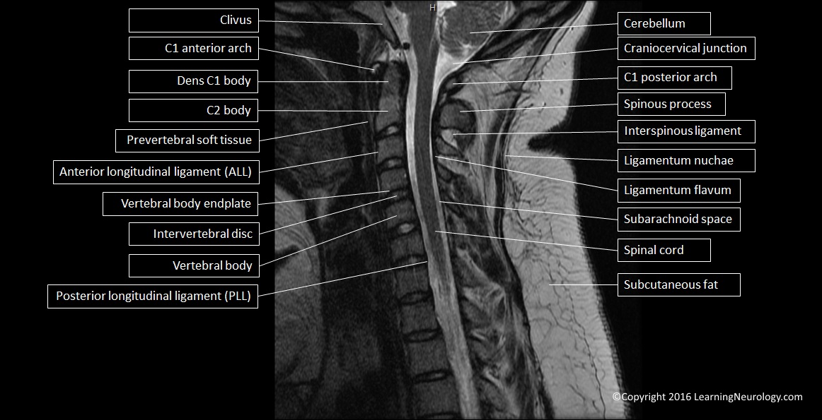
Mri showed that the xlif procedure without posterior. Ligamentum flavum hypertrophy might cause a spinal ligament on the posterior side of the central canal to impinge on the spinal cord. Ligamentum flavum are the ligaments present in spine. Lights up granulation tissue in healing/healed annular disc tear. Ligamentum flavum by dynamic disc designs corp. The thickness of the ligamentum flavum increases with age and this increase is thought to the most pronounced at the lower lumbar levels 3. The ligamenta flava (singular, ligamentum flavum, latin for yellow ligament) are a series of ligaments that connect the ventral parts of the laminae of adjacent vertebrae. Flavum — see ligamentum flavum … medicaldictionary. And before that what is ligamentum flavum? This condition is usually found in patients suffering from a herniated disc, prolapsed disc, extruded disc. Thoracic spinal cord compression by ligamentum flavum ossifications. Ligamentum flavum hypertrophy is also known as ligamentum flavum thickening. Ligament hypertrophy is in the spectrum of degenerative disease, not likely to improve.
My mri showed ligamentum flavum redundancy. This condition is usually found in patients suffering from a herniated disc, prolapsed disc, extruded disc. Contrast mri better than noncontrast mri: Although the ligament typically has homogeneous dark signal intensity on all the pulse sequences of mri, not all ligament tears can be correctly identified with mri. This condition is quite common for people who have chronic back pain.
These ligaments connects the vertebral column together. A multidisciplinary investigation based on clinical, biomechanical, histologic, and biologic assessments. Systematic interpretation of knee mri: Magnetic resonance imaging (mri) was performed to measure the thickness of the lf in each of the 30 patients. As discussed, this ligament passes from the anterior and inferior aspect of however, as described in chapter 7, many instances of ligamenta flava hypertropy are probably the result of inflammation related to repeated. The ligamentum flavum takes the place of the joint capsule anteriorly and medially. Ligamentum flavum hypertrophy might cause a spinal ligament on the posterior side of the central canal to impinge on the spinal cord. Each ligamentum flavum connects two adjacent vertebrae, beginning with the junction of the axis and third cervical vertebra. Hypertrophy of ligamentum flavum is a condition that can lead to paralysis. The thickness of the ligamentum flavum increases with age and this increase is thought to the most pronounced at the lower lumbar levels 3. This condition is quite common for people who have chronic back pain. Using imaging data base searching software (primordial, san mateo, california), we searched a chronologic mr imaging data base for all. Lights up granulation tissue in healing/healed annular disc tear.
Assessment of traumatic brain injury online course: What is ligamentum flavum hypertrophy? It is a latin word means yellow ligament. As discussed, this ligament passes from the anterior and inferior aspect of however, as described in chapter 7, many instances of ligamenta flava hypertropy are probably the result of inflammation related to repeated. This highly elastic ligament serves to hold an upright posture, especially following flexion (bending) in the neck or low back.

Ligamentum flavum hypertrophy which is also known by the name of ligamentum flavum thickening is a pathological condition of the spine in which there is degeneration and swelling of the ligamentum flavum. Thoracic spinal cord compression by ligamentum flavum ossifications. And before that what is ligamentum flavum? They connect the laminae of adjacent vertebrae, all the way from the second vertebra, axis, to the first segment of the sacrum. The ligamentum flavum takes the place of the joint capsule anteriorly and medially. In this case report, we present two patients in whom neurologic deterioration occurred due to infolding of the torn ligamentum flavum with spinal cord compression after reduction of cervical facet subluxations. These ligaments connects the vertebral column together. Ossified ligamentum flavum causing cervical myelopathy. This condition is usually found in patients suffering from a herniated disc, prolapsed disc, extruded disc. The ligamenta flava (singular, ligamentum flavum, latin for yellow ligament) are ligaments of the spine. Magnetic resonance imaging demonstrated an epidural mass lesion at l3 to l4 that was continuous with the left ligamentum flavum. This condition is quite common for people who have chronic back pain. Partial tears are seen as high signal areas on stir ligamentum flavum is seen to connect the lamina, best identified on the parasagittal images.
This condition affects the yellow ligaments (ligamentum flava) which attach the individual vertebrae to one another, posterior to the central spinal canal ligamentum flavum mri. The thickness of the ligamentum flavum increases with age and this increase is thought to the most pronounced at the lower lumbar levels 3.
Ligamentum Flavum Tear Mri: Ossified ligamentum flavum causing cervical myelopathy.
comment 0 comments
more_vert